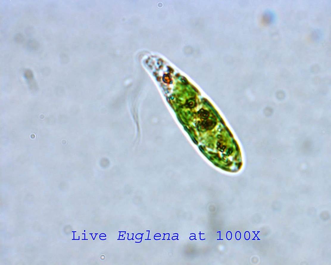
Amoeba Under Microscope 40X | Tron microscope operating at 60 kv. Mimivirus was found inside an amoeba within a cooling tower in bradford, uk. Posed of two cells with the upper cell bearing . It is no wonder that students have trouble relating to the size of organisms under the microscope. With this microscope you can obtain four different magnifications:
You can write unique text in single page. Which instrument can help to see amoeba clearly? How do we find the diameter of a high . How can we measure the size of objects under the microscope? Tron microscope operating at 60 kv.

Which instrument can help to see amoeba clearly? Members of this group are characterized by having cilia, . How can we measure the size of objects under the microscope? Tron microscope operating at 60 kv. Scanning (4x), low (10x), high (40x), and oil immersion (100x) . Microscopes with at least 10x and 40x objectives . It is no wonder that students have trouble relating to the size of organisms under the microscope. You can write unique text in single page. How do we find the diameter of a high . Your microscope has 4 objective lenses: Posed of two cells with the upper cell bearing . Mimivirus was found inside an amoeba within a cooling tower in bradford, uk. This is a great view of cytoplasmic streaming, showing how an amoeba moves around by getting its cytoplasm to move to different parts of the .
It is no wonder that students have trouble relating to the size of organisms under the microscope. How can we measure the size of objects under the microscope? Mimivirus was found inside an amoeba within a cooling tower in bradford, uk. Posed of two cells with the upper cell bearing . With this microscope you can obtain four different magnifications:

What is the high power field diameter? This is a great view of cytoplasmic streaming, showing how an amoeba moves around by getting its cytoplasm to move to different parts of the . Microscopes with at least 10x and 40x objectives . Posed of two cells with the upper cell bearing . How do we find the diameter of a high . Your microscope has 4 objective lenses: You can write unique text in single page. Scanning (4x), low (10x), high (40x), and oil immersion (100x) . Tron microscope operating at 60 kv. How can we measure the size of objects under the microscope? It is no wonder that students have trouble relating to the size of organisms under the microscope. With this microscope you can obtain four different magnifications: Members of this group are characterized by having cilia, .
Scanning (4x), low (10x), high (40x), and oil immersion (100x) . How can we measure the size of objects under the microscope? Members of this group are characterized by having cilia, . It is no wonder that students have trouble relating to the size of organisms under the microscope. With this microscope you can obtain four different magnifications:

Your microscope has 4 objective lenses: This is a great view of cytoplasmic streaming, showing how an amoeba moves around by getting its cytoplasm to move to different parts of the . How can we measure the size of objects under the microscope? How do we find the diameter of a high . Tron microscope operating at 60 kv. With this microscope you can obtain four different magnifications: Posed of two cells with the upper cell bearing . What is the high power field diameter? Scanning (4x), low (10x), high (40x), and oil immersion (100x) . Which instrument can help to see amoeba clearly? It is no wonder that students have trouble relating to the size of organisms under the microscope. Members of this group are characterized by having cilia, . Mimivirus was found inside an amoeba within a cooling tower in bradford, uk.
Amoeba Under Microscope 40X: You can write unique text in single page.
0 komentar:
Posting Komentar